
Figure 4.
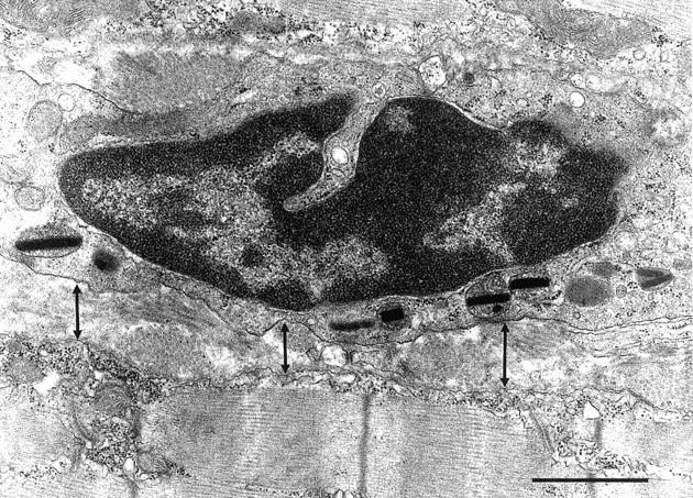
Electron micrograph of 4-week-old mdx muscle showing an eosinophil lying within the endomysial connective tissue that separates two muscle fibers that show no signs of pathology. The eosinophil is separated from the muscle fiber surface by ∼0.5 μm (arrows) and the MBP rods within the eosinophil are oriented along the length of the eosinophil. Scale bar, 1.0 μm.