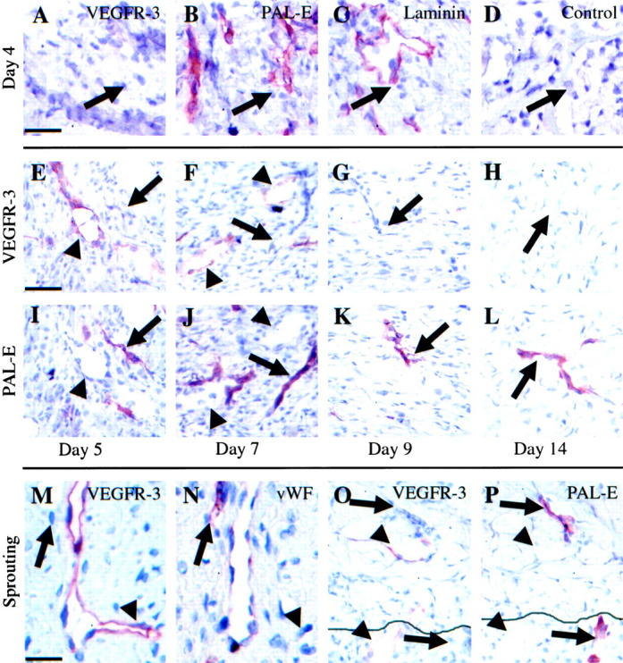
Figure 2.

Analysis of VEGFR-3-positive vessels and blood vessels in wound healing. The angiogenic vessels at the leading edge on day 4 stained with VEGFR-3 (A), PAL-E (B), laminin (C), or control antibodies (D). Granulation tissue on days 5, 7, 9, and 14 stained for VEGFR-3 (E−H) and PAL-E (I−L). Note that the VEGFR-3-positive vessels (arrowheads) do not stain for PAL-E (arrows) and vice versa. Sprouting of the VEGFR-3-positive vessel in granulation tissue of a day 7 punch biopsy wound stained for VEGFR-3 (M) and vWF (N). Note absence of VEGFR-3 on blood vessel endothelium (arrows) and the very slight staining of vWF in lymphatic endothelium. New lymphatic vessel (arrowheads) and blood vessel (arrows) sprouting from pre-existing vessels at the edge of a day 5 punch biopsy wound stained for VEGFR-3 (O) and PAL-E (P). The continuous line in O and P marks the wound edge. Scale bars, 50 μm (A−D, M, N) and 70 μm (E−L, O, P).
