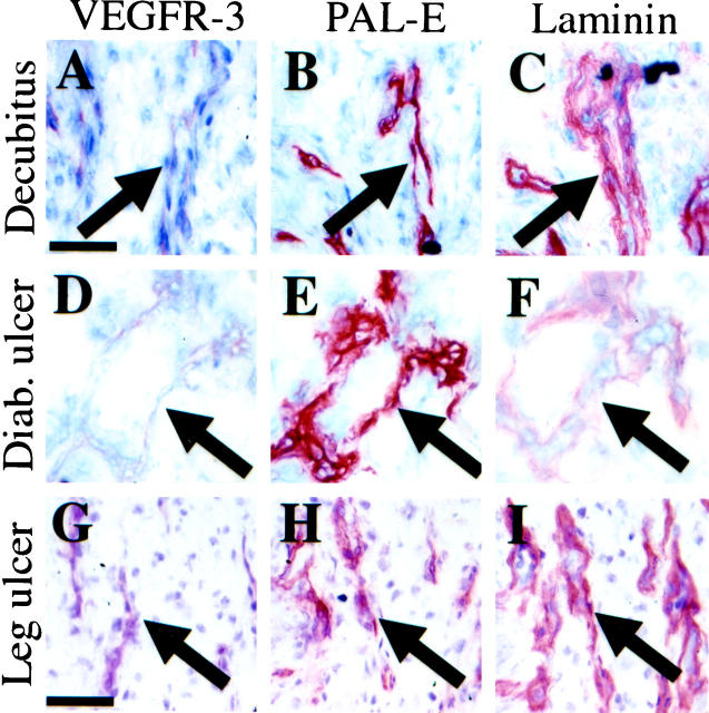
Figure 4.
Immunoperoxidase staining of serial sections of a decubitus lesion (A−C), a diabetic leg ulcer (D−F), and a nondiabetic leg ulcer (G−I). Blood vessels (arrows) stain very weakly for VEGFR-3 (A, D, and G) and strongly for PAL-E (B, E, and H) and laminin (C, F, and I). Scale bars, 100 μm (A−F) and 200 μm (G−I).

