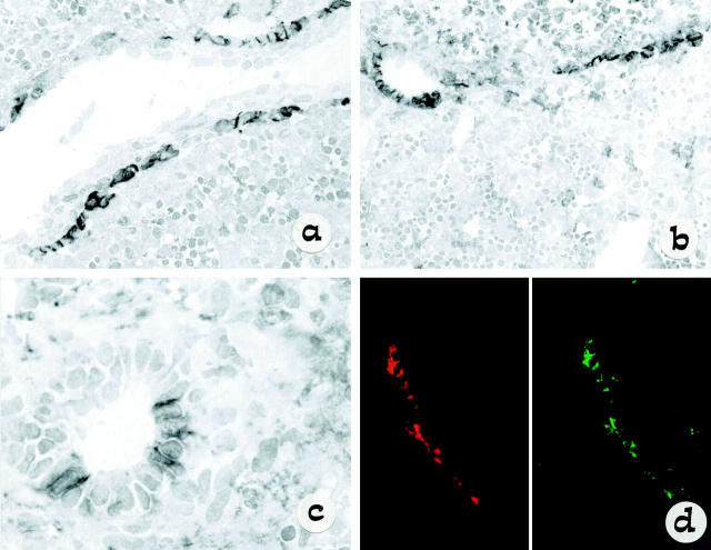
Figure 1.
Light micrograph of single staining (immunoperoxidase method) showing the distribution of NCAM during the early steps in bile duct morphogenesis (a–c). An indirect three-step immunoperoxidase technique was used with peroxidase-labeled rabbit anti-mouse and swine anti-rabbit immunoglobulin (Dako) and diaminobenzidine substrate. NCAM is expressed on a single-layer ductal plate surrounding a large portal vein branch, as seen in fetal liver at 12 weeks’ gestation (a: original magnification, ×60). However, NCAM is disappearing as duplicated segments are formed with staining still present in residual ductal plate remnants, as seen in fetal liver at 14 weeks’ gestation (b: original magnification, ×40). Faint patchy NCAM staining is observed in bile ducts with an easily recognizable lumen incorporating into mesenchyma, as seen in fetal liver at 16 weeks’ gestation (c: original magnification, ×120). In d the high level of colocalization between NCAM (red) and Bcl-2 (green) is demonstrated in developing ductal plate by dismerged images of double staining at confocal analysis.

