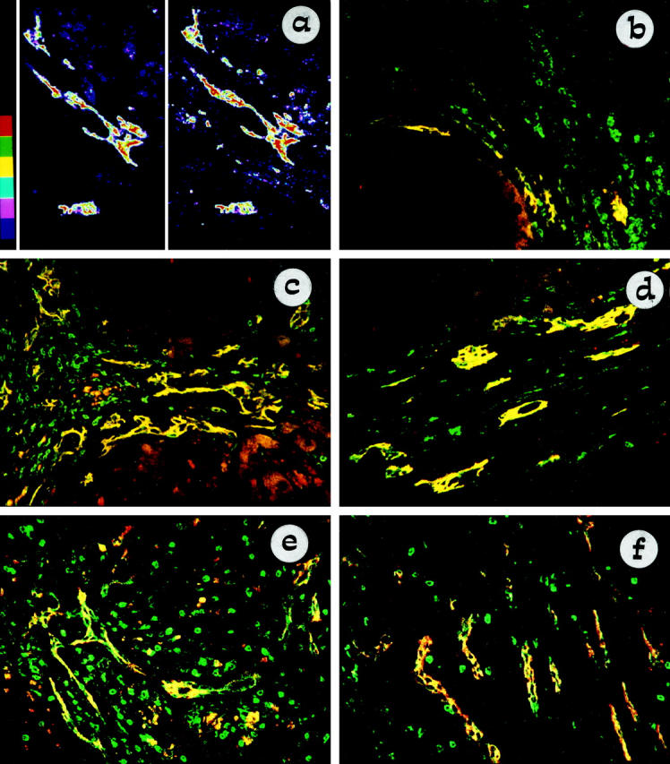
Figure 3.

Confocal micrograph of double staining with NCAM (Texas Red) and Bcl-2 (FITC), showing coincident expression between these two markers on atypical ductules. In a the level of coincidence between NCAM (left side) and Bcl-2 (right side) is analyzed by dismerging and color banding in a biliary atresia tissue sample: the highest intensities are shown in red, and the lowest intensities are in blue. NCAM and Bcl-2 share the same distribution and concentration on biliary epithelium: the highest and the lowest levels of binding of NCAM and Bcl-2 are localized at the same places within the atypical ductular structures. All control reactions demonstrated the specificity of the double staining. In b–f, NCAM and Bcl-2 coexpression on ductular reaction is reported for different chronic hepatobiliary diseases: cryptogenic cirrhosis (b), PSC (c), biliary atresia (d), polycystic liver (e), and Caroli’s disease (f). Areas of coincident labeling appear yellow. Bcl-2 noncoincident staining (green) is observed in lymphocytes. In parenchymal liver cirrhosis (b) only a small subset of reactive ductules, characterized by a marginal distribution, coexpressed NCAM and Bcl-2. In primary cholangiopathies (c and d) and developmental liver diseases related to ductal plate malformation (e and f), coincident immunoreactivity for NCAM and Bcl-2 was extensively observed among atypical ductules. In c granular NCAM staining (red) was found in scattered periseptal hepatocytes.
