
Figure 5.
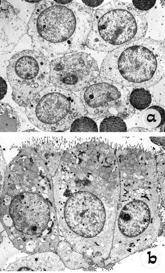
Ultrastructural analysis by TEM of cells freshly isolated from Percoll gradients by immunomagnetic separation, using anti-NCAM (a) and HEA-125 (b) on the NCAM-depleted population. Note the polar columnar morphology, with basal nuclei and well-formed apical microvilli of mature biliary epithelial cells (b), compared with the rounded shape, high nucleus-cytoplasmic ratio, short scattered microvilli, and fewer junctional complexes of NCAM-positive cells (a).