
Figure 1.
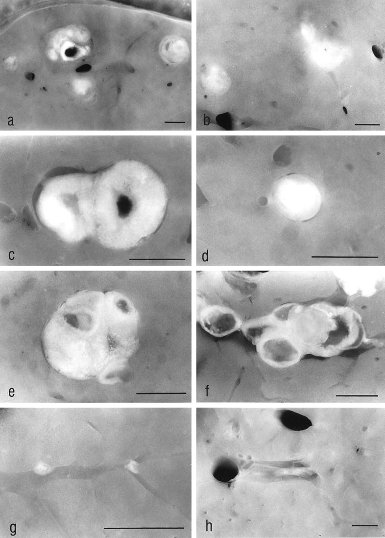
The stereomicroscopic examination of unstained liver slices allowed us to find intrahepatic ovarian transplants. In the ovariectomized animals (MG) (a and c) and in the orchiectomized animals (CG3) (b), the transplants reached a size of about 2 mm at 3 weeks after transplantation, whereas the transplants in the nonovariectomized animals (CG2) remained small (d). Three months after transplantation the transplants of MG (e), as well as those of the toremifene-treated animals (CG5) (f), showed a similar size if compared with those at 3 weeks after transplantation, whereas the transplants of CG2 (g) and of CG3 (h) underwent fibrosis, obviously because they were not stimulated in a sufficient manner by gonadotropins. The altered liver tissue downstream of the transplants in MG cannot be identified in the unstained slices. The transplant shown in e is the same as shown in Figure 5a ▶ . a–d: 3 weeks after transplantation; e–h: 3 months after transplantation. Original magnifications: a, ×9; b, ×11; c, ×25; d, ×29; e, ×22; f, ×18; g, ×35; h, ×11. Scale bars, 1 mm