
Figure 4.
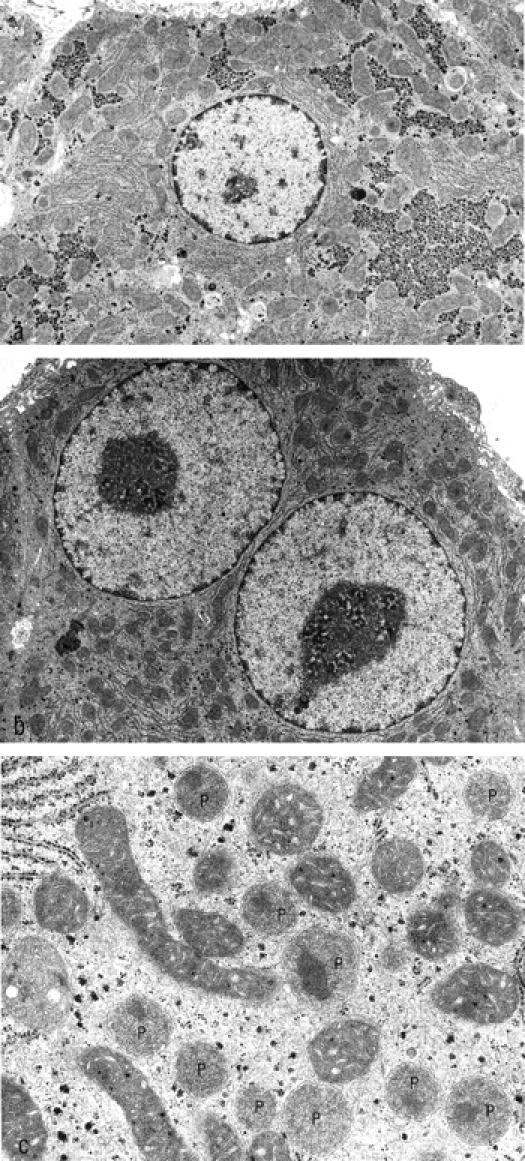
The electron micrographs show nonaltered (a) and altered (b) hepatocytes of the same animal 3 weeks after transplantation. The altered hepatocytes exhibit a loss of glycogen and enlarged nuclei with a decondensation of chromatin and prominent nucleoli. At higher magnification (c), a strong increase in the number and size of peroxisomes can be identified (P). Original magnifications: a and b, ×7,850; c, 32,600.