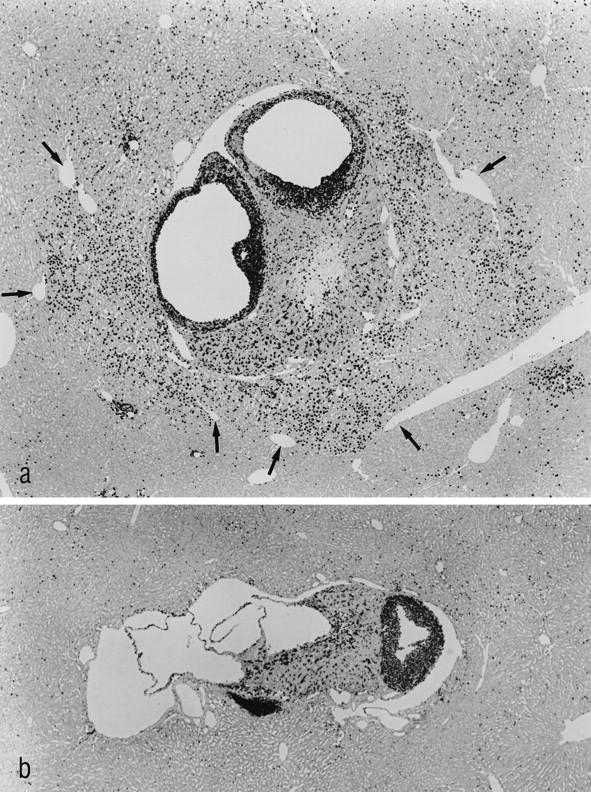
Figure 5.

The immunohistochemical localization of BrdU-labeled nuclei demonstrates a strong proliferative activity of the transplant and of the surrounding hepatocytes in MG 3 months after transplantation (a). The border of the liver acini downstream of the transplants is sharply defined by the draining hepatic venules (arrows), demonstrating the acinar nature of the hyperproliferative areas. b: The transplant of a toremifene-treated animal 3 months after transplantation is found to proliferate, whereas the surrounding hepatocytes do not show any alteration in their proliferative activity. Shown are immunostains for BrdU (administered via s.c. implanted osmotic minipumps for 7 days). Original magnification: a and b, ×48.
