
Figure 7.
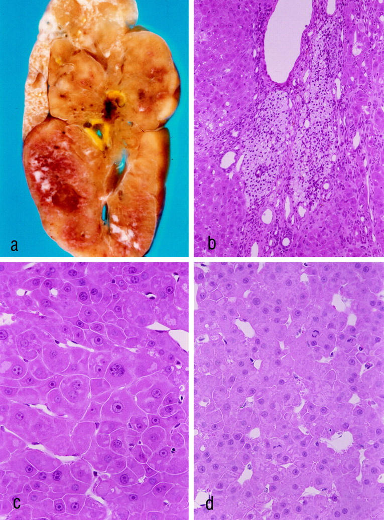
Hepatocellular carcinoma (HCC) 18 months after ovarian tissue transplantation (a–d). a–c: The same HCC, which measures 4 cm in diameter. At this time after transplantation all transplants consisted of luteinized theca cells. b: The transplant is surrounded by the HCC. c and d: The HCCs show a trabecular growth pattern. The tumor cells share some characteristics with the altered hepatocytes at 3 weeks and at 3 months after transplantation (compare with Figure 3 ▶ , b–d), ie, large nucleoli, cytoplasmic amphophilia, and sometimes small areas of glycogen accumulation. Mitotic figures are visible in d. a: Unstained liver slice after perfusion fixation. b–d: H&E stains. Original magnifications: a, ×3; b, ×180; c and d, ×360.