
Figure 4.
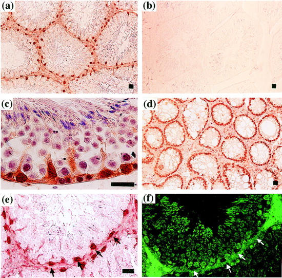
Localization of Rbm3 protein expression in mouse testis. Immunohistochemical staining of the testes of 4-month-old C57BL/6 mice (a, b, c, e, and f) and W/Wv mutant mice deficient in germ cells (d). Sections were incubated with the rabbit anti-Rbm3 antibody (a, c, d, and e) or preimmune antiserum (b), and bound antibody was detected using the anti-rabbit IgG conjugated with horseradish peroxidase. f: A serial section of (e) was incubated with the TRA 98 germ cell-specific antibody, 24 and bound antibody was detected using the fluorescein isothiocyanate-conjugated secondary antibody and fluorescence microscopy. Arrowheads indicate the Rbm3-positive Sertoli cells. Scale bar, 20 μm.