
Figure 5.
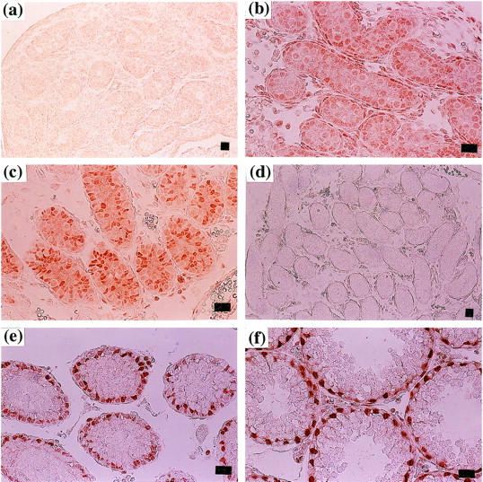
Expression of Rbm3 during testicular development. Testes were isolated from 17 days postcoitus fetus (a), newborn (b), 8-day (c and d), 17-day (e), and 21-day (f) -old mice. Immunohistochemical staining was performed using the anti-Rbm3 antibody (a, b, c, e, and f) or preimmune antiserum (d). Scale bar, 20 μm.