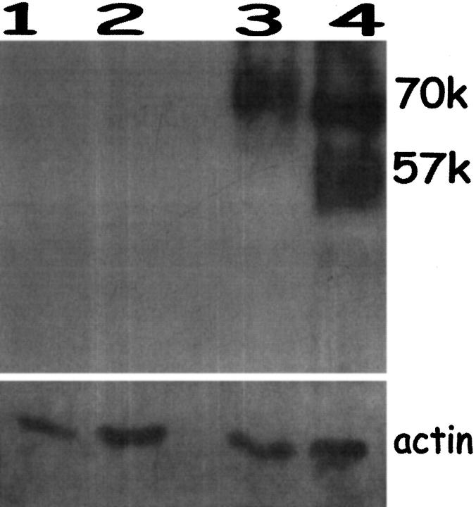
Figure 5.
Immunoblotting for EPO in endometriosis lesions and endometrium from endometriosis patients. Lanes 1 and 2: normal endometrium from two endometriosis patients, proliferative phase. Lanes 3 and 4: endometriosis tissue from proliferative phase. Only the endometriosis samples contained abundant EPO. Size markers were included. Reprobing of these blots for actin indicated equal loading of protein in all lanes.

