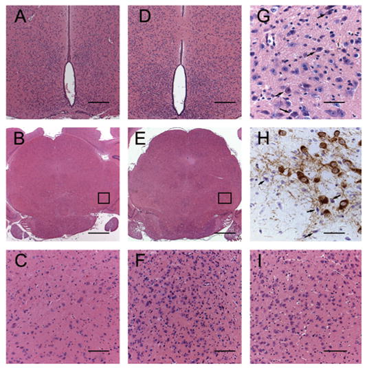
Figure 2.

The normal morphology of dorsomedial and ventromedial hypothalamic nuclei in a 9-wk-old wild-type male mouse (A) is readily distinguished, when compared with the increased cellular infiltrate within the periventricular hypothalamic nuclei of an 8-wk-old homozygous recessive male mouse (D). The increased number of cells in this region is approximately doubled in the homozygous recessive mouse and obscures the normal periventricular morphology (D). The pars compacta and pars reticulata have a moderate to marked cellular infiltrate in PKU female mice at 8 wk (E and F) and occurs as early as 4 wk (I). The SN of a 9-wk-old wild-type female (B and C) is within normal limits. High-power magnification reveals numerous round cells (arrows, G) that are 8–10 μm in diameter with amphophilic cytoplasm, round nuclei, deeply basophilic homogeneous chromatin pattern, and inconspicuous nucleoli. These cells are pictured in the SN of 8-wk-old PKU mice. Infiltrative cells are present within the pars compacta of a male PKU mouse adjacent to DA neurons (TH; arrows, H). The infiltrative cells present in the pars compacta of PKU mice occasionally feature TH immunoreactive cytoplasmic granules. (A and D) H&E; bar = 100 μm. (C and F) H&E; bar = 50 μm. (G) H&E; bar = 25 μm. (H) TH immunoperoxidase method; bar = 25 μm.
