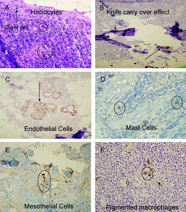
FIGURE 1. False-positive IHC staining in sentinel lymph nodes. A, H&E stained section of SLN in a patient that underwent biopsy of colon adenocarcinoma; typical postbiopsy changes of giant cells (arrow), and hemosiderin-laden histiocytes (circles). B, cytokeratin IHC section demonstrating extraneous matter and transferred tumor cells (arrow), knife carry-over effect. C, immunohistochemical endothelial cell staining (arrow). D, characteristic positive IHC staining of endogenous peroxidase-rich granular mast cells. E, positively staining mesothelium (circle) in omental fat fragment adherent to SLN on IHC. F, typical histologic appearance of nodal pigmented macrophages (circle).
