
Figure 3.
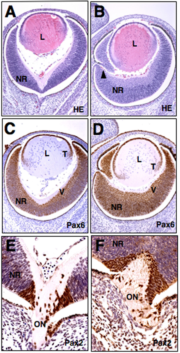
Retinal coloboma in Rybp+/- mouse embryo. (A-B) Hematoxylin and eosin- stained coronal sections of normal (A) and Rybp heterozygous null (B) eyes at E14.5. The neuroretina of the mutant eye is thickened and fails to close leading to the formation of coloboma (B; arrowhead). (C-D) Immunolocalization of Pax6 in wild type (C) and mutant (D) eyes. Pax6 is normally expressed in the ventral side of neuroretina. In the mutant eyes it shows broader expression in the retina and it is also more posteriorly positioned in the transition zone of the lens (C compare to D). (E-F) Immunolocalization of Pax2 in wild type (E) and mutant (F) eyes. L; lens, NR; neuroretina, ON; optic nerve, V; ventral, T; transitional zone. HE; hematoxylin and eosin. Magnifications: A-D(×320); E-F(×460)