Figure 9.
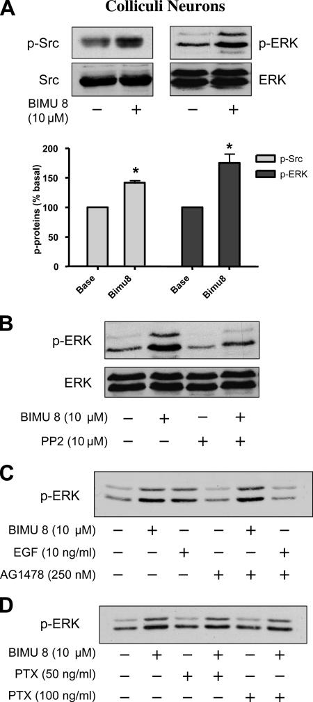
Src-dependent 5-HT4R–mediated ERK1/2 activation also occurs in neurons. (A) Colliculi neurons endogenously expressing 5-HT4R were stimulated with vehicle or BIMU8 (a 5-HT4R agonist). Total neuron lysates were analyzed by immunoblotting with the indicated antibodies. The same immunoblot was probed sequentially for p-Src with antibody to active p-Src (Tyr 416), for total inactive Src with antibody against pan Src, for p-ERK1/2 with the antibody to activate ERK1/2, and for total ERK1/2 with the antibody to ERK42/44. Quantification of p-Src and p-ERK1/2 was performed by densitometric analysis using NIH Image software. Data are means ± SEM of results obtained in four independent experiments. *p < 0.05 versus corresponding values measured in untreated neurons. (B) Colliculi neurons were pretreated with the Src kinase inhibitors PP2 at 10 μM for 30 min before the stimulation with 10 μM BIMU8 for 5 min. Neuron lysates were analyzed by immunoblotting with antibodies against p-Src and p-ERK1/2 as indicated in A. (C) 5-HT4R–mediated ERK activation does not require transactivation of EGF-R tyrosine kinase. Colliculi neurons were pretreated with 250 nM tyrphostin/AG1478 for 30 min before stimulation with 10 μM BIMU8 or 10 ng/ml EGF during 5 min. Total lysates were analyzed by immunoblotting with antibody to p-ERK1/2. (D) 5-HT4R–mediated ERK activation does not require G protein PTX sensitive (Gi/Go). Colliculi were pretreated with PTX at 50 or 100 ng/ml overnight before treatment with 10 μM BIMU8 for 5 min. Total lysates were analyzed by immunoblotting with antibody to p-ERK1/2. A representative blot of each experiment is shown in A–D.

