Figure 5.
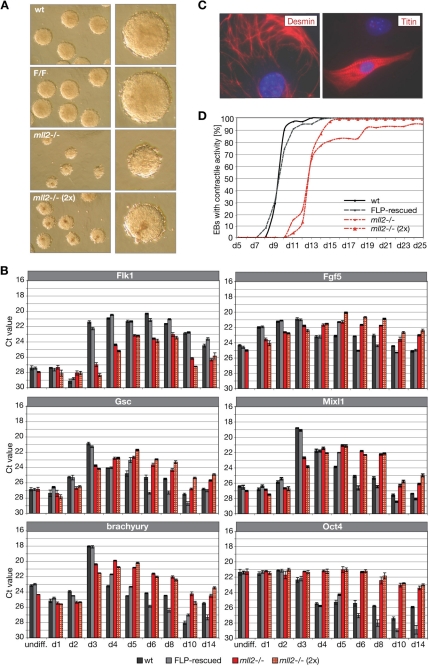
Cardiac differentiation of ES cells in vitro. (A) Embryoid body (EB) formation of wild-type, F/F, and mll2−/− ES cells on day 3 of cardiac in vitro differentiation, magnification 20× and close-up. EBs designated as mll2−/− (2x) were generated from 2 times more cells than the other three panels. (B) Quantitative expression analysis of early differentiation markers Flk1 (early mesendoderm), Gsc and Mixl1 (gastrulation), Fgf5 (epiblast), brachyury (mesoderm), and the pluripotency marker Oct4 upon cardiac in vitro differentiation. Total RNA from EBs at different time points was isolated and processed for reverse transcription. Ct values of qPCR analysis were normalized against ribosomal protein L19 (RPL19). Data are means of triplicate analysis ± SD. (C) Regions of cardiogenesis were identified by appearance of cardiac specific markers. Contracting regions of mll2 mutant ES cells were dissected, dissociated, and processed for cardiac-specific immunoreactivity by using anti-desmin and anti-titin antibodies. Magnification, 40×. (D) Appearance of spontaneous contractile activity during EB outgrowth. EBs, derived from a defined number of cells (1 × 10³ cells/EB), were plated and checked for appearance of contractile activity (until day 25). In addition, mll2−/− EBs resulting from 2 times more cells (2 × 10³ cells/EB) were monitored. Data were obtained from the same experimental series, n = 96 EBs.

