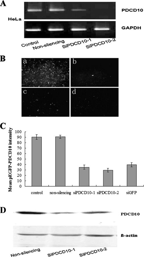
Figure 4.
Transfection of siPDCD10-1 or siPDCD10-2 inhibited PDCD10 expression at both mRNA and protein levels. (A) RT-PCR PDCD10 mRNA expression data. PC-3 cells, which express high levels of PDCD10, were transfected with nonsilencing siRNA or the indicated siRNAs against PDCD10. At 24 h after transfection, total RNAs were extracted and RT-PCR experiments were performed as described (see Materials and Methods). Both siPDCD10-1 and siPDCD10-2 showed much stronger inhibitory effects for PDCD10 mRNA expression than nonsilencing siRNA. (B) Effects of siRNAs against PDCD10 on the expression of GFP PDCD10 fusion protein. PC-3 and HeLa cells were cotransfected with pEGFP-PDCD10 plasmid and siPDCD10-1 (c) and siPDCD10-2 (d), respectively. Plasmids encoding pEGFP-PDCD10 were also transfected into PC-3 and HeLa cells cotransfected with nonsilencing siRNA (a) as negative control, as well as siRNA against pEGFP (b) as positive control. At 24 h after transfection, cells were observed by fluorescence microscopy. siPDCD10-1 and -2–transfected cells show far fainter fluorescence than cells transfected with nonsilencing siRNA. Then cells were harvested and immediately analyzed by FACS Calibur (1 × 10⁴ cells) and CELLQuest software (BD Bioscience). Mean GFP-PDCD10 fluorescence intensity was quantificationally shown in C. (D) Immunoblot assessment on the effects of siPDCD10 on endogenous PDCD10 protein expression. HeLa cells were transfected with nonsilencing siRNA or siPDCD10. At 24 h after transfection, a significant decrease in the expression of endogenous PDCD10 was observed in siPDCD10–transfected cells, whereas no decrease was noted in nonsilencing siRNA transfected cells. β-actin was used as the internal control.

