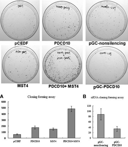
Figure 6.
PDCD10 modified normal and MST4-induced cellular growth in colony forming assays. (A) Left two panels, PC-3 cells transfected with pCDEF-PDCD10, pCDEF-MST4, pCDEF-PDCD10, and pCDEF-MST4 or vector control in monolayer culture and selected with G418. Quantitative analyses of colony numbers are shown in the right panel (n = 3). (B) Representative sample of inhibition of colony formation in monolayer culture by siPDCD10. Right panel, PC-3 cells transfected with pGC-PDCD10, or nonsilencing control in monolayer culture. Quantitative analyses of colony numbers are shown in the right panel.

