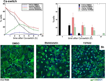
Figure 4.
ROCK-mediated myosin II inhibition promotes transient lateral lumina in Ca2+-switch assays. MDCK cells were incubated with DMSO (1:2000), 50 μM blebbistatin, or 20 μM Y-27632 for 30 min in SMEM before and during Ca2+ switch. Merged confocal x-y views of gp135 and ZO-1 5 h after Ca2+ switch. Note the higher incidence of lateral lumina in blebbistatin- and Y-27632–treated monolayers. Graph, percentage of cells with VACs and lateral lumina in DMSO and blebbistatin-treated cells (left) and DMSO- and Y-27632–treated cells (right). Bars, 10 μm.

