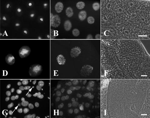
Figure 7.
Overexpression of GFP-Nopp140-True (D–F) and GFP-Nopp140-RGG (G–I) in salivary glands leads to disrupted nucleolar (nuclear) structure and lethality. (A–C) Third instar larvae homozygous for the second chromosome insertion, pUAST-GFP-Nopp140-True.A9, were heat-shocked for 1 h and allowed to recover for 2 h. (A) Moderate expressions of GFP-Nopp140-True from the Hsp70 promoter failed to perturb nucleolar or nuclear morphology. (B) DAPI staining showed the polytenic chromatin distributed throughout the nuclei. (C) Phase-contrast microscopy showed the phase-dark nucleoli. (D–F) pUAST-GFP-Nopp140-True.A9/+; +/Sgs3-GAL4 third instar larvae overexpressed GFP-Nopp140-True in salivary glands. (D) Nucleoli appeared swollen and disorganized. (E) DAPI-stained chromatin. (F) Nucleoli were not readily discernible by phase-contrast microscopy. (G–I) pUAST-GFP-Nopp140-RGG.G4/+; +/da-GAL4 progeny overexpressed GFP-Nopp140-RGG that filled the nuclear volume. Most nucleoli appeared disrupted by fluorescence and phase-contrast microscopy. Arrows show a few nucleoli that remained normal in size. Most progeny died in the larval stages, but a few died as pupae. Bars, 100 μm

