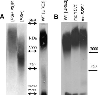
Figure 2.
Agarose gel electrophoresis of prion polymers. Particulate fractions from sucrose pad centrifugation analyzed on 1.5% agarose gels and blotted to nitrocellulose. Immunostaining was with anti-Sup35 or anti-HA antibody. (A) Size comparison of [PSI+PS], [PSI+], and [URE3]. (B) Size differences of [URE3] prion polymers for wild-type cells and cells overproducing (mc) either Ydj1p or Sse1p.

