Figure 8.
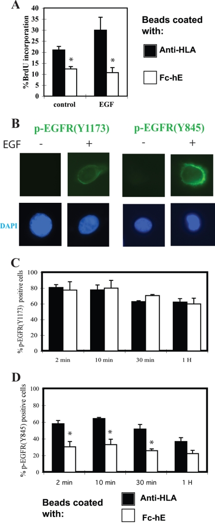
E-cadherin ligation selectively inhibits EGF receptor Tyr845 phosphorylation. (A) E-cadherin ligation inhibits EGF-induced increase in growth of A431 cells. A431 cells plated in low serum (0.5%) were incubated with 10 ng/ml EGF, and beads were coated with either anti-HLA or Fc-hE for 24 h. The percentage of cells entering S phase was determined by the cell proliferation assay. (B) EGF treatment induces the immunofluorescence staining of phosphorylated-EGFR (Tyr1173) and phosphorylated-EGFR (Tyr845) in A431 cells. DAPI staining of nuclei is shown at bottom. (C) E-cadherin ligation has no effect on EGFR autophosphorylation at Tyr1173. A431 cells were serum starved and incubated with either anti-HLA or Fc-hE–coated beads overnight, and then they were stimulated with 10 ng/ml EGF for the indicated times and stained with antibody to phosphorylated-EGFR (Tyr1173). The number of p-EGFR (Tyr1173)-positive cells was counted and percentage of positive cells was calculated. (D) E-cadherin ligation selectively inhibited EGFR Tyr845 phosphorylation. A431 cells were treated as in C and stained with antibody to phosphorylated-EGFR (Tyr845). The number of p-EGFR (Tyr845)-positive cells was counted, and percentage of positive cells was calculated. Data are expressed as mean ± SEM (*p < 0.05).

