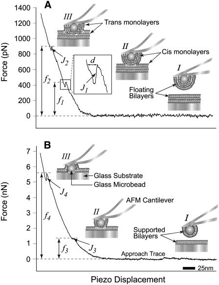
FIGURE 1.
Illustrations (I–III (A and B); not to scale) and typical AFM force scans (force versus piezo-displacement) in the presence of floating (A) and supported (B) lipid bilayers showing the jumps (J1-4) and the associated fusion forces (f1-4). Force scans in panels A and B show that initially no interaction is taking place between the bilayers at long distances (I) and therefore the force is equal to zero. Upon further approach, the cantilever is bent as the bilayers start to press against one another and the force starts to increase. With the continued application of force, the bilayers are compressed together until fusion takes place (II) and a first jump (J1 or J3) is observed at ∼450 pN (f1) or ∼1.4 nN (f3), respectively. The jump is the result of the sudden movement of the cantilever tip toward the substrate as fusion of the floating bilayers (A) or supported bilayers (B) takes place (see illustrations). The piezo-electric transducer displacement (d, inset) during the jump is a measure of its distance and reflects the thickness of the fused bilayer. A transient reduction in force takes place as the cantilever tip relaxes during the jump, followed by an increase upon further bending with the continued application of compression, which eventually leads to the appearance of a second jump (J2 or J4) at ∼900 pN (f2) or ∼5.5 nN (f4), which is indicative of the fusion of the remaining floating bilayer (A) or supported (B), respectively (III).

