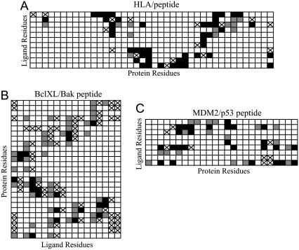
FIGURE 2.
Residue-residue contacts for the three ligand-protein complexes used to evaluate docking methods. Residue-residue interactions present in both the x-ray crystal structures and in the closest docked models are shown in black. False negatives are shown with a cross, and false positives are shown in gray.

