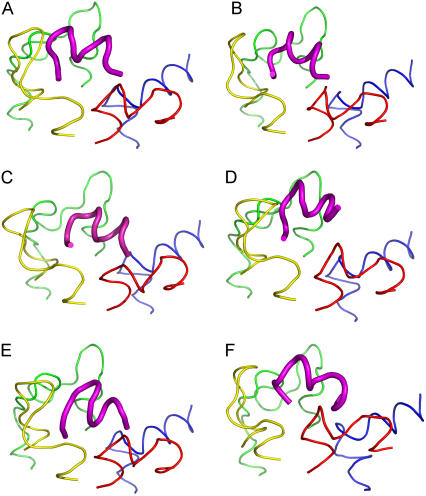
FIGURE 3.
(A) Gtα(340-350) and its analogs. (B) Peptide 2, (C) Peptide 11, (D) Peptide 14, (E) Peptide 3, and (F) [R341, S347]-Gtα(340-350) are shown in the common binding mode. The first and last three Cα atoms in the loop structures were superimposed to find the common binding pose. IC1 is shown in red, IC2 in yellow, IC3 in green and IC4 in blue. Gtα(340-350) and its analogs are shown in magenta. The figure was rendered in PYMOL (DeLano Scientific, Palo Alto, CA).

