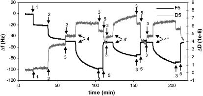
FIGURE 3.
QCM-D measurements of the binding reactions outlined in Fig. 1. Immobilization of SA (0.1 mg/ml) and ERE (200 nM) was done in PBS buffer, and binding of proteins was carried out in HEPES buffer containing 100 mM KCl. The initial change in both f and D observed upon addition of ERα samples includes not just protein adsorption but also a buffer change effect (the ERα working solution contains some glycerol absent in the baseline HEPES buffer). Upon rinsing the surface at the end of the protein binding, this buffer effect is removed and the endpoint Δf and ΔD are recorded. The surface with immobilized ERE is regenerated using 0.1% SDS to remove bound proteins and the baseline is reset with HEPES buffer.

