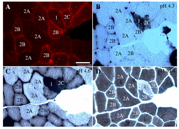
Figure 3.

Immunohistochemistry of β-synemin and ATPase staining on muscle sections after injection with cardiotoxin. (A) 10 μm transverse muscle sections were immunostained with an antibody against β-synemin. (B-D) ATPase staining on serial sections was performed at pH 4.3 (B), pH 4.6 (C), and pH 10.4 (D). Type 1 fibers (labeled with a "1") show strong staining at pH 4.3 and pH 4.6 and no staining at pH 10.4, whereas type 2A and 2B fibers (labeled with a "2A" and "2B") show the reverse. Type 2A fibers show weaker staining than type 2B at pH 4.3 and pH 4.6. In addition, type 2C fibers reacted at pH 4.3, pH 4.6, and pH 10.4 and are under the process of regeneration. Bar = 50 μm
