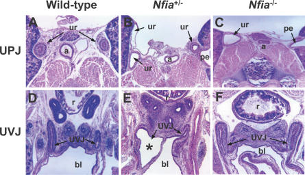
Figure 7. Ureter Defects at UPJ and UVJ in Nfia Mutant Mice.
(A–C) Hematoxylin and eosin histology depicts duplication and dilatation of ureter at the UPJ in Nfia +/− (B, 60X) and Nfia −/− (C, 60X) newborn mice.
(D–F) Hematoxylin and eosin staining shows dilatation of UVJ in some Nfia +/− mutant mice (* in E, 60X); the majority of Nfia +/− and Nfia −/− mice show a normal UVJ (F, 60X).
a, abdominal aorta; bl, bladder; pe, pelvis; r, rectum; ur, ureter; UVJ, ureterovesical junction

