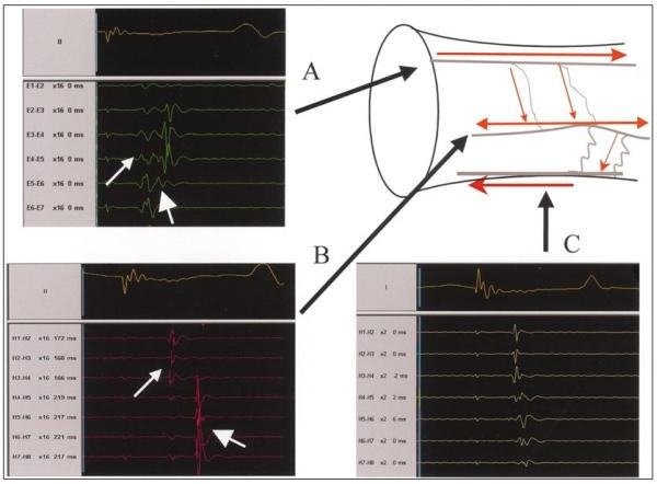
Figure 5.

Illustration of longitudinal and transverse patterns of spline activations. A; A spline with longitudinal activation. The earliest PV activity is recorded at electrode pair E 5-6. B; Transverse pattern of activation, manifested as simultaneous recordings along a spline. The simultaneous recording of PV potentials along a spline suggests that the electrodes along the spline are activated by a wide electrical wavefront propagating perpendicular to several electrodes along the spline. C; Transverse activation that is simultaneous on the most distal end (H4-5 through H1-2), but with activation from distal to proximal (from H4-5 through H7-8). When a narrow wavefront traveled initially along the long axis of the vein and later spread perpendicular to the long axis of the vein, it might reach a spline from its distal end first, then travel along a fiber leading to a sequence of activation from distal to proximal. Reprinted with permission from Sanchez, JE et al, Evidence for longitudinal and transverse fiber conduction in human pulmonary veins: relevance for catheter ablation. Circulation 2003; 108: 590-597.
