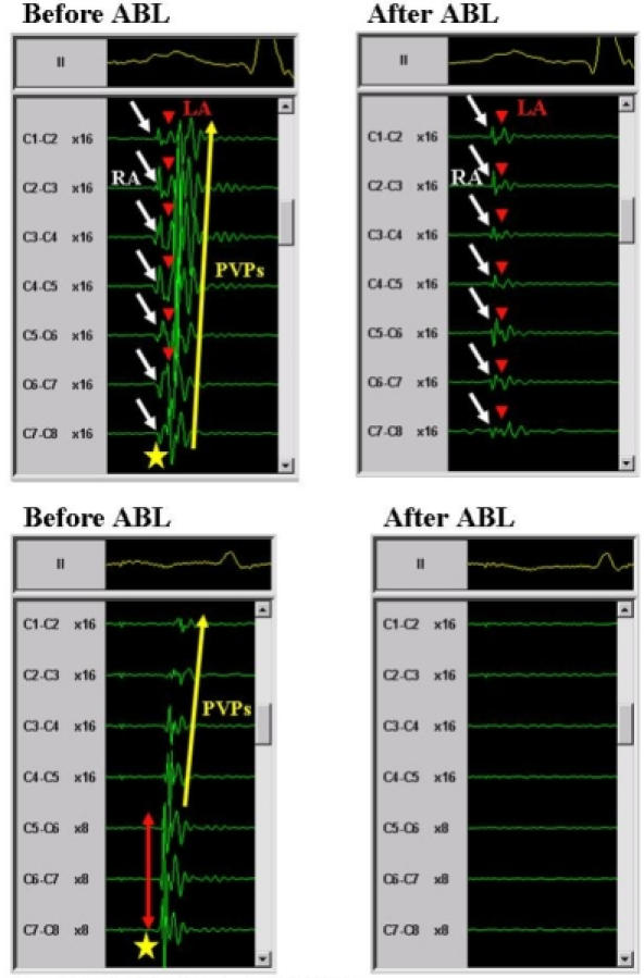
Figure 7.

Definition of PV antrum potentials. A upper panels; An antrum potential (star) recorded from the right superior PV during sinus rhythm (left panel). Note that the antrum potential was a single sharp potential, which was formed by the total fusion of the PV potentials (PVPs) and left atrial (LA) potentials around the PV ostium. After the PV antrum ablation, all the PVPs and PV antrum potentials disappeared and only the right atrial (RA) and LA potentials were observed (right panel). The white arrows indicate the RA potentials, red arrowheads the LA potentials and yellow arrow the PVPs. B lower panels; An antrum potential (star) recorded from the LSPV during distal CS pacing (left panel). Note that the antrum potential had single sharp potentials, which exhibited a transverse activation pattern around the PV ostium. The red arrow indicates the transverse activation pattern and yellow arrow the PVPs. After the PV antrum ablation, all the PVPs and PV antrum potentials disappeared and no potentials were observed (right panel). The abbreviations are as in figure 6. Reprinted from Yamada T et al. Electrophysiological pulmonary vein antrum isolation with a multielectrode basket catheter is feasible and effective for curing paroxysmal atrial fibrillation: efficacy of minimally extensive pulmonary vein isolation. Heart Rhythm 2006;3:377-384. Copyright (2006), with permission from The Heart Rhythm Society.
