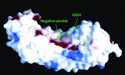
Fig. 3.
grasp (39) electrostatic potential surface of PGIP2. Regions of negative and positive potential are shown in red and blue, respectively. A wide negative pocket, putatively involved in PG recognition, is located in the middle of the inner concave surface of the protein. The residue Gln-224, crucial for PGIP2 specificity, is also indicated.

