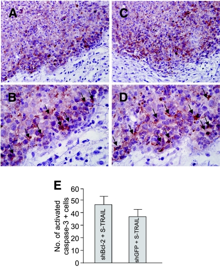
Figure 6.
Immunohistochemistry detects activated caspase-3 in glioma cells. Mice implanted with a mix of LV-S-TRAIL or control vector-transduced and nontransduced Gli36-EGFRvIII-Fl-shGFP or Gli36-EGFRvIII-Fl-shBcl-2 cells (Figure 5) were sacrificed on day 6 after tumor cell implantation, and tumors were sectioned and stained with anti-caspase-3 antibodies. The stained sections were counterstained with hematoxylin. (A and B) Sections from S-TRAIL-expressing Gli36-EGFRvIII-Fl-shGFP gliomas. (C and D) S-TRAIL-expressing Gli36-EGFRvIII-Fl-shBcl-2 gliomas. Caspase-3-stained cells are shown by arrows. (E) The number of activated caspase-3 positive cells was calculated by counting the positive cells in randomly selected field of views under a microscope. Original magnification, ×10 (A and C) and ×40 (B and D).

