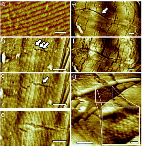
Fig. 1.
Disintegration of the spore coat rodlet layer. (a) The intact rodlet layer covering the outer coat of dormant B. atrophaeus spores is ≈11 nm thick, and has a periodicity of ≈8 nm (14). (b–d) Series of AFM height images tracking the initial changes of the rodlet layer after 13 min (b), 113 min (c), and 295 min (d) of exposure to germination solution. Small etch pits (indicated with arrows in b) evolve into fissures (indicated with an arrow in c) perpendicular to the rodlet direction. The fissures expand both in length and width. (e and f) Series of AFM images showing another germinating spore. The spore long axis, as well as major rodlet orientation, is left-right. Enhanced etching at stacking faults (running from left to center and indicated with an arrow in e), as well as increased etching at the perpendicular fissures, was visible after 135 min (e) and 240 min (f) of germination. Fissure width and length increased from 10–15 nm and 100–200 nm (135 min) to 15–30 nm and 125–250 nm (240 min), respectively. (g) Etching and/or fracture of the rodlet layer at a stacking fault revealed the underlying hexagonal layer of particles with a 10- to 13-nm lattice period. [Scale bars: 20 nm (a and g Inset) and 100 nm (b–g).]

