Fig. 4.
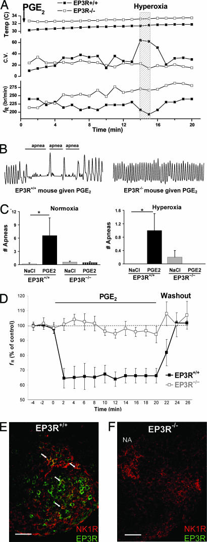
PGE2 depresses brainstem respiratory activity and induces apnea via brainstem EP3Rs. Respiration was examined in EP3R+/+ (n = 13) and EP3R−/− (n = 25) neonatal mice after administration of PGE2 (n = 19) or NaCl (n = 19). (A) PGE2 was injected i.c.v. at 0 min followed by normoxia and a 1-min hyperoxic challenge in newborn EP3R+/+ (■) and EP3R−/− (□) mice. The EP3R+/+ mouse exhibited a lower fR (breaths per min) and an irregular respiratory rhythm with elevated coefficient of variation (C.V.) during normoxia and hyperoxia due to apneic breathing. In the EP3R−/− mouse, basal fR did not decrease after the postanesthesia period, and there was less variability in the respiratory pattern. No temperature difference or dependency was observed during the first 20 min after i.c.v. administration of PGE2. (B) Plethysmograph recordings (10-s periods with breath amplitude of 1 μl/s) demonstrate apnea episodes in response to PGE2 during normoxia in an EP3R+/+ mouse, but not in an EP3R−/− mouse. (C) In EP3R+/+ mice, PGE2 induced more apneas during normoxia and hyperoxia compared with vehicle. This effect of PGE2 was not observed in EP3R−/− mice. (D) In en bloc brainstem spinal–cord preparations from 2- to 3-d-old EP3R+/+ pups (■, n = 5), PGE2 (20μg/liter) reversibly depressed respiratory rhythm generation to 64 ± 5% of control frequency (fR) (ANOVA repeated measures design, P < 0.01). PGE2 did not affect respiratory activity in preparations from EP3R−/− mice (□, n = 6). (E) In transverse medullary sections, respiration-related neurons within the RVLM ventral to the nucleus ambiguus (NA) and including the preBötC coexpress NK1R (red) and EP3R (green). The arrows indicate EP3R and NK1R colocalization (yellow) in some RVLM respiration-related neurons. (F) NK1R, but no EP3R, expression was identified in an EP3R−/− mouse. (Scale bar, 100 μm.) Data are presented as mean ± SEM. ∗, P < 0.05 compared with EP3R+/+ mice given NaCl.

