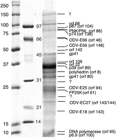
Fig. 1.
ODV proteins were separated on a 7–15% gradient gel and stained with Coomassie blue. The bands were subjected to in-gel trypsin, and their determined identity is listed to the right. Those bands marked with a question mark either did not produce significant peptides for analyses, or the peptide masses did not match predicted peptides from the AcMNPV genome. Prestained and unstained standards are used routinely; however, they vary significantly in the lower molecular weight range. As such both standards are included for reference [prestained, far left (Bio-Rad; precision); unstained, center (Bio-Rad, LMW)].

