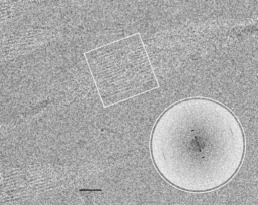
Fig. 4.
Wild-type FBP28 filaments at ≈5 mg/ml unstained and frozen in amorphous ice. Imaged by using cryoelectron microscopy (cryo-em) at 1.8 μm underfocus (protein is dark), this single twist is a 180° rotation of the ribbon's projected density. The dense crossovers are clear, but the 2.5-nm rods of density are visible only close to where the ribbon is face-on. There is a low-density region within each rod. (Scale bar, 20 nm.) (Inset) A calculated Fourier transform of the boxed area. Across the ribbon are two orders of 2.5-nm spacing, whereas there is an 0.47-nm spacing along the ribbon.

