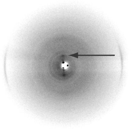
Fig. 5.
X-ray diffraction of wild-type FBP28 fibers (≈10 mg/ml) after washing into water and drying down onto film. The x-ray pattern was taken in the plane of the film and shows the 0.47-nm arcs typical of cross-β structure found in amyloid filaments. A weak equatorial spacing is also seen at ≈2.8 nm (arrow).

