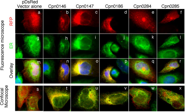
Figure 5.
Localization of RFP-Cpn fusion proteins in host cell endoplasmic reticulum (ER). HeLa cells transfected with pDsRed plasmids encoding various C. pneumoniae proteins as listed on top of the figure were subjected to immunofluorescence staining. The RFP or RFP fusion proteins were in red while host cell endoplasmic reticulum was labeled with a rabbit antibody against Calnexin (as an ER marker) in combination with a goat anti-rabbit IgG conjugated with Cy2 (green) and DNA labeled with Hoechst dye (blue). The slides were observed under both conventional fluorescence (panels a-r) and confocal (s-x) microscopes. Note that the microscopic observations revealed ER co-localization of Cpn0146 (panels n & t), 0147 (o & u) & 0186 (IncA; p & v) but not RFP alone (m & s), Cpn0284 (q & w) or 0285 (r & x).

