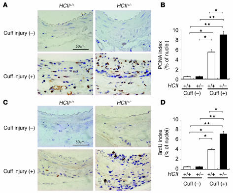
Figure 6. Immunostaining of proliferative vascular mesenchymal cells with PCNA and BrdU at the vascular wall with and without cuff injury in HCII+/+ and HCII+/– mice.
(A) PCNA staining of the vascular wall with and without cuff injury in HCII+/+ and HCII+/– mice. (B) Quantitative analysis of PCNA-positive cells in intima and media with and without cuff injury in HCII+/+ (white bars) and HCII+/– mice (black bars). Values are expressed as mean ± SEM. *P < 0.05; **P < 0.01. n = 8 in each group. (C) BrdU staining of the vascular wall with and without cuff injury in HCII+/+ and HCII+/– mice. (D) Quantitative analysis of BrdU-positive cells in intima and media with and without cuff injury in HCII+/+ (white bars) and HCII+/– mice (black bars) Values are expressed as mean ± SEM. *P < 0.05; **P < 0.01. n = 8 in each group.

