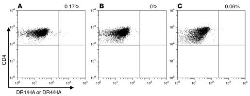
Figure 4. Representative tetramer staining of DRB1*0101+ cord blood stained with the DR1/HA tetramer (A), the same DRB1*0101+ cord blood stained with the DR4/HA negative control tetramer (B), and a sample from a DRB1*0101– adult volunteer stained with the DR1/HA tetramer (C).
Panels show percentage of tetramer-staining–positive cells from a representative subject.

