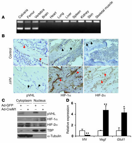
Figure 2. Osteoblast-specific, Cre-mediated deletion of Vhl.
(A) PCR analysis of Cre-mediated recombination in selected tissues from a ΔVhl mouse. The recombined allele (Δflox) was present exclusively in bone tissue. (B) Representative histological sections of distal femurs from 6-week-old control and ΔVhl mice after staining with antibodies against pVHL (left), HIF-1α (middle), or HIF-2α (right) as described in Methods. Sections were counterstained with hematoxylin. Red arrows indicate positive staining and black arrows negative staining in osteoblasts. Original magnification, ×400. (C and D) Confluent monolayers of Vhl floxed primary osteoblasts were infected with either Ad-GFP or Ad-CreM1 (100 MOI) for 48 hours. (C) Proteins in the cytoplasm and nucleus were extracted separately and analyzed by immunoblotting with antibodies against pVHL, HIF-1α, and HIF-2α. Immunoblots for TBP and α-tubulin were used as loading controls for nuclear and cytoplasmic proteins, respectively. TBP, TATA box–binding protein. (D) Total mRNA was extracted from confluent monolayers of osteoblasts 48 hours after adenoviral infection, and gene expression for Vhl, Vegf, and Glut1 was determined by quantitative real-time PCR. White bars represent Ad-GFP infection; black bars represent Ad-CreM1 infection. *P < 0.05; **P < 0.01.

