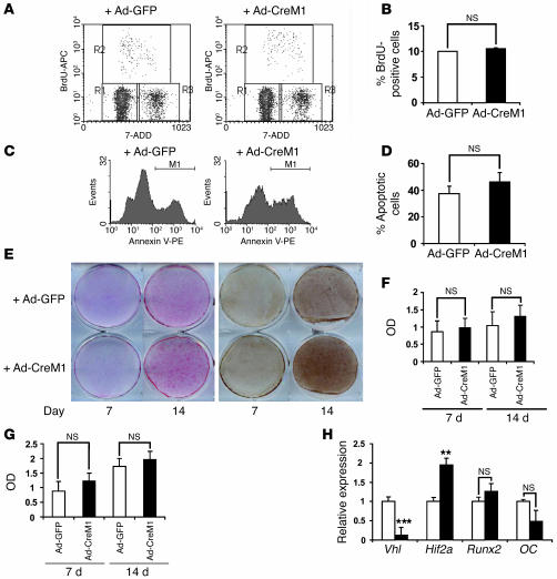
Figure 6. Deletion of Vhl in primary osteoblasts in vitro does not affect osteoblast proliferation and apoptosis.
Confluent Vhl floxed primary osteoblast monolayers were infected with either adeno-GFP or adeno-CreM1 (100 MOI). Vhl mRNA expression in infected osteoblasts was determined by real-time PCR 48 hours after infection to assess deletion efficiency. Cell proliferation, apoptosis, and differentiation assays were performed as described in Methods. (A and B) Cell proliferation was assessed by flow cytometry using BrdU incorporation. (C and D) Cell apoptosis was assessed by flow cytometry using annexin V–PE staining. (E) Mineralized nodule formation was determined by ALP (left) and von Kossa staining (right) 7 and 14 days after cells were cultured in osteogenic medium. (F and G) Densitometric analysis of ALP and von Kossa staining observed in E using NIH ImageJ 1.36b. Data represent mean ± SEM. (H) Measurement of Vhl, Hif1a, runt-related transcription factor 2 (Runx2), and OC mRNA expression by quantitative real-time PCR at day 14 of osteogenic induction. **P < 0.01; ***P < 0.001.

