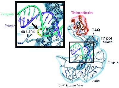
Figure 1.
Structural alignment of T7 and Taq DNA polymerases. Atomic coordinates of the crystal structures of Taq DNA polymerase (38) (PDB entry 2KTQ) and T7 DNA polymerase (35) (PDB entry 1T7P) were superimposed by using the program insight ii (Biosym Technologies, San Diego). The superimposed structures were visualized by using the program setor (41). T7 DNA polymerase (light grey) bound to the thioredoxin molecule (orange), primer (purple), and the template (cyan) are shown. The different domains of the T7 DNA polymerase are indicated. Only the I and I1 helices of Taq DNA polymerase (dark grey) are shown here, superimposed on the corresponding helices of T7 DNA polymerase. The loop unique to T7 DNA polymerase, comprising residues Glu-Gly-Asp-Lys spanning from residue 401 through 404, is shown in green.

