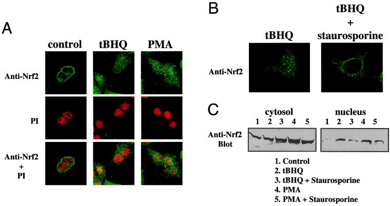
Figure 2.
Involvement of PKC in the nuclear localization of Nrf2. (A) HepG2 cells were treated with DMSO (control), tBHQ (100 μM), or PMA (100 nM) for 4 h. Nrf2 localization was detected by immunocytochemistry using an anti-Nrf2 antibody followed by FITC-conjugated secondary antibody. To confirm the location of the nuclei, PI counterstaining of the same fields was shown both separately and overlaid with Nrf2 immunofluorescence. (B) HepG2 cells were preincubated with solvent or staurosporine (15 nM) for 1 h before their exposure to tBHQ (100 μM) for 4 h. Nrf2 immunofluorescence was monitored as in A. (C) Subcellular fractionation of HepG2 cells was carried out after treatment of tBHQ (100 μM) or PMA (100 nM) for 1 h, in the presence or absence of staurosporine (15 nM). Eighty micrograms of proteins of the cytosolic fraction and 20 μg of the nuclear fraction were resolved by SDS/PAGE. The relative amounts of Nrf2 protein present in each were determined by Western blotting using an anti-Nrf2 antibody followed by enhanced chemiluminescence detection.

