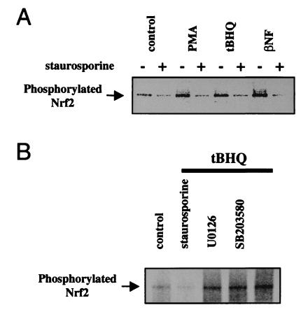
Figure 4.
Involvement of PKC in Nrf2 phosphorylation in vivo. 32P-labeling of HepG2 cells was followed by immunoprecipitation of Nrf2 as in Fig. 3. (A) Cells were treated with tBHQ (100 μM), βNF (50 μM), or PMA (100 nM) for 30 min, after preincubation for 1 h in solvent or in 15 nM staurosporine. (B) Cells were exposed to tBHQ (100 μM) for 30 min, after preincubation for 1 h in solvent, staurosporine (15 nM), U0126 (10 mM), or SB203580 (10 μM).

