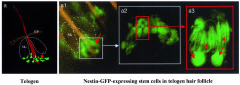
Fig. 2.
Hair follicle nestin-GFP-expressing cells in the telogen phase of nestin-GFP transgenic mouse skin. The skin sample was prepared freshly right after excision from the back skin of a nestin-GFP transgenic mouse. The skin sample then was directly observed by fluorescence or confocal microscopy with the dermis side up after s.c. tissue was dissected out. (a) Cartoon of telogen hair follicle showing position of nestin-GFP-expressing hair follicle stem cells. (a1) Low-magnification fluorescence microscopy image showing the ring of bulge nestin-GFP-expressing stem cells (small white box, see a). (a2) High-magnification confocal microscopy image reflecting the small white box in a1. Note the small round- or oval-shaped nestin-GFP-expressing cells in the bulge area of the hair follicle (small red box). (a3) High-magnification fluorescence microscopy image showing two individual nestin-GFP-expressing stem cells reflecting the red box in a2. Note the unique morphology of the hair follicle stem cells and multiple dendrite-like structures of each cell. Red arrows indicate the cell body, and red arrowheads show the multiple dendritic structure of each cell. (Original magnifications: a1, ×100; a2, ×400; a3, ×1,600.) SG, sebaceous gland; EP, epidermis.

