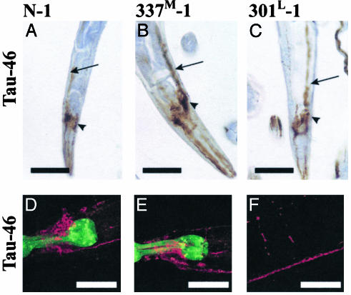
Fig. 4.
Immunostaining for Tau. (A–C) Paraffin-embedded sections were stained with antibody Tau-46. (D–F) Whole mount animals were immunostained (37) by using Tau-46 as the primary antibody and AlexA 568 as the secondary antibody (red). GFP in the pharynx is green. Arrows point to the ventral nerve cord, and arrowheads are head neurons. No staining was seen in non-Tg animals (not shown). D and E, head; F, commissures, dorsal, and ventral nerve cords (see also Fig. 10, which is published as supporting information on the PNAS web site). (Scale bars = 50 μm.)

