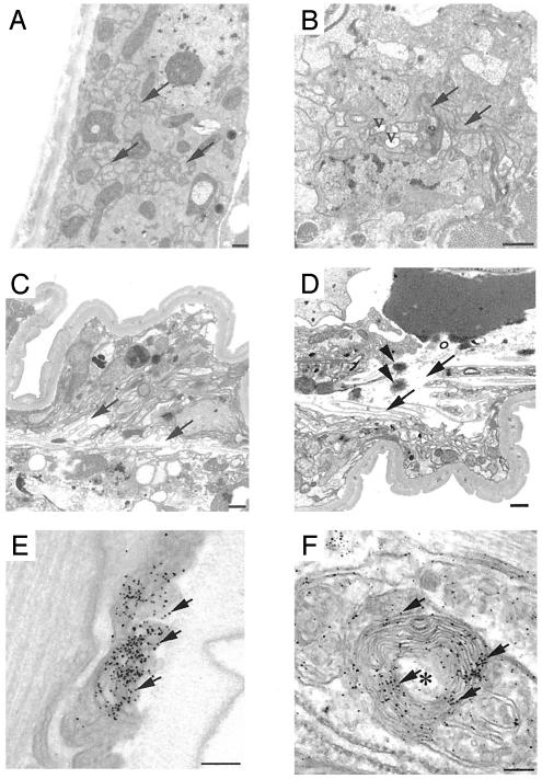
Fig. 6.
Ultrastructural of degenerating axons. (A–D) Transmission EM. Transverse sections of 9-day-old N-1 (A), 301L-1 (B), and 337M-1 (C and D) lines. Arrows show normal axons in A, mildly degenerative axons in B, and severely degenerating disoriented axons in C and D. V, vacuolar clearing of axoplasm. Spherical, osmophilic protein aggregates (arrowheads) were evident (D). (E and F) Preembedding immuno-EM of 337M-1 animals by using antibody 17026. The arrow shows immuno-silver-enhanced particles that identify tau-labeled structures. Degenerating axons with collapsed membranous profiles (onion skin lesions) are indicated by an asterisk and contain amorphous aggregates labeled by anti-tau antibodies. (Scale bars = 500 nm in A–D and 100 nm in E and F.)

